

Advanced dental imaging technology improves diagnosis by providing high-resolution, three-dimensional views of hard and soft tissues that traditional 2D X-rays cannot capture. This clarity allows dentists to detect early-stage decay, assess bone density, and plan complex procedures with greater precision. At LI Dental Group in Garden City, NY, we utilize these modern tools to ensure accurate treatment planning and better patient outcomes.
The Shift from 2D to 3D Imaging
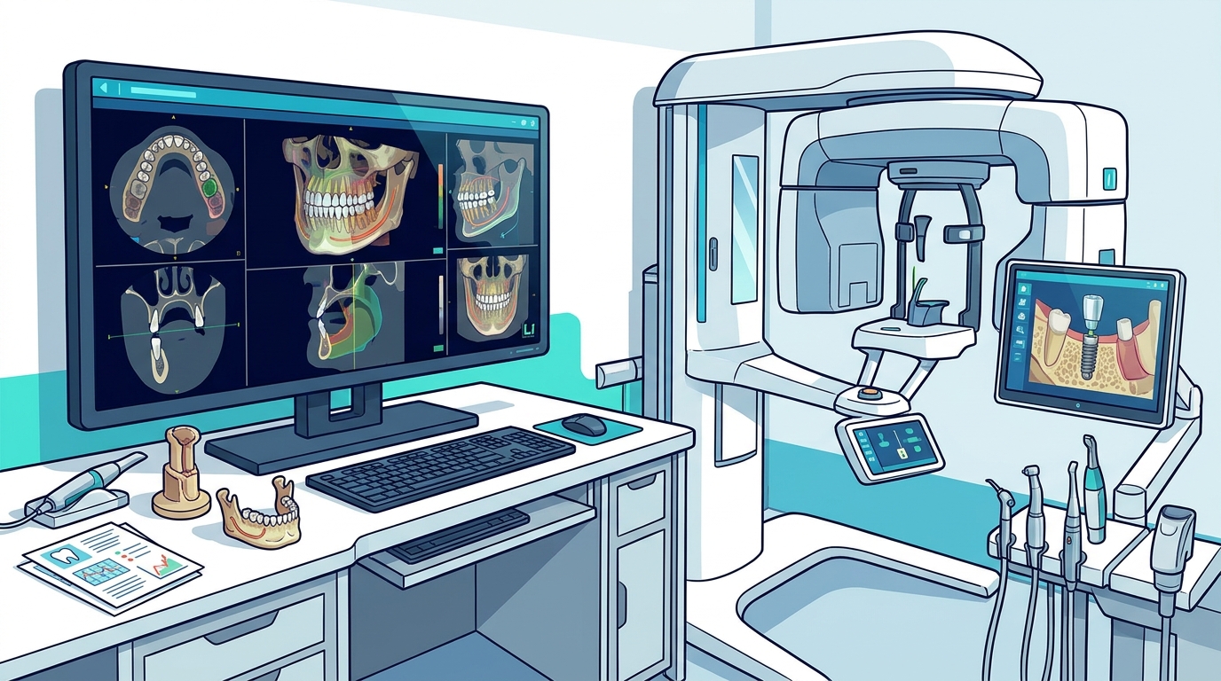
Traditional dental radiography relies on two-dimensional images that flatten complex anatomical structures into a single plane. This limitation can obscure critical details, such as the exact depth of a cavity or the proximity of a nerve to a root. Cone-beam computed tomography (CBCT) is a type of 3D imaging that captures detailed cross-sectional views of the jaw, teeth, and surrounding tissues. By moving beyond flat images, clinicians can visualize the spatial relationships between structures that were previously hidden.
Recent literature reviews highlight that the transition from 2D to 3D imaging represents a crucial change in dental diagnostics. This shift advances patient care through improved accuracy and comprehensive treatment planning. For example, when planning a dental implant, a 3D scan reveals the exact volume and quality of the jawbone. This information is vital for determining if a bone graft is necessary before the implant can be placed. Without this depth of data, treatment plans may lack the precision required for long-term success.
Artificial Intelligence in Diagnostic Imaging
Modern imaging systems are increasingly integrated with artificial intelligence (AI) to enhance diagnostic capabilities. Artificial intelligence in dentistry is the use of algorithms and machine learning models to analyze medical images and assist in clinical decision-making. These systems can process large volumes of data quickly, identifying patterns that might be missed by the human eye. AI tools can annotate radiographs, highlight areas of potential pathology, and even predict treatment outcomes based on historical data. according to Artificial Intelligence in
Research indicates that AI assistance improves diagnostic efficiency and interobserver agreement while reducing interpretation time. For instance, AI-based software can analyze patient data and case histories to assist dentists in creating personalized treatment plans. In a clinical setting, this means faster identification of issues like early gum disease or hidden fractures. At LI Dental Group, we leverage these technological advancements to ensure that no subtle sign of oral health issues goes unnoticed during your exam. according to Advancements in Diagnostic
Precision in Treatment Planning
Accurate diagnosis is only half the benefit of advanced imaging; the other half is precise treatment planning. When a dentist has a 3D map of the oral cavity, they can simulate procedures before they begin. This is particularly important for complex cases involving full-arch implant reconstruction or orthodontic alignment. Digital treatment planning is the process of using computer software to design and visualize a patient's dental restoration before any physical work is done.
For patients considering Invisalign, 3D imaging allows the dentist to map out the exact sequence of tooth movements required to achieve the desired result. This level of detail ensures that the final smile aligns with both aesthetic goals and functional needs. Similarly, for restorative procedures like crowns or bridges, digital scans eliminate the need for traditional putty impressions. Instead, a digital scanner captures the exact contours of the tooth, leading to restorations that fit more precisely and comfortably. This precision reduces the number of adjustments needed and speeds up the overall treatment process.
Enhancing Patient Comfort and Safety
Advanced imaging technology also contributes to patient comfort and safety. Digital X-rays and 3D scanners use significantly less radiation than older film-based systems. The FDA and the American Dental Association have established guidelines to ensure that radiation exposure is kept to the minimum necessary for diagnosis. By using modern equipment, practices can adhere to these safety standards while providing superior diagnostic quality.
Furthermore, the speed and non-invasive nature of digital scanning reduce patient anxiety. Many patients dread the discomfort of traditional impressions or the uncertainty of waiting for X-ray results. With digital tools, the process is faster, cleaner, and more transparent. Patients can often view their own scans on a screen, which helps them understand their condition and the proposed treatment. This transparency builds trust and empowers patients to make informed decisions about their oral health. At LI Dental Group, we prioritize these patient-centered technologies to create a stress-free and informative experience.
Key Takeaways
- 3D imaging provides detailed views of jawbone and soft tissues that 2D X-rays cannot show.
- AI integration helps dentists identify pathology faster and with greater consistency.
- Digital treatment planning allows for the simulation of procedures before they begin.
- Modern imaging systems use less radiation than traditional film-based methods.
- Patients benefit from faster, cleaner, and more transparent diagnostic processes.
- Advanced technology supports precise implant placement and orthodontic alignment.
Frequently Asked Questions
Is 3D dental imaging safe?
Yes, 3D dental imaging is considered safe. It uses low-dose radiation, and the benefits of accurate diagnosis generally outweigh the minimal risks. Practices follow strict guidelines to minimize exposure.
How does AI help in dental diagnosis?
AI analyzes images to detect patterns and anomalies that may be difficult for humans to see. It assists dentists in identifying issues like early decay or bone loss more efficiently.
Do I need a 3D scan for every visit?
No, 3D scans are typically reserved for specific cases, such as implant planning or complex orthodontics. Routine checkups usually require standard 2D X-rays.
What is the difference between a digital X-ray and a 3D scan?
A digital X-ray provides a flat, 2D image, while a 3D scan creates a detailed, volumetric model of the jaw and teeth, allowing for deeper analysis.
Can advanced imaging detect gum disease?
Yes, advanced imaging can assess the bone levels supporting the teeth, which is a key indicator of the severity of gum disease.
Experience Precision Care at LI Dental Group
Advanced dental imaging technology is transforming how we diagnose and treat oral health issues. By combining 3D imaging with AI-driven analysis, we can offer you more accurate, comfortable, and efficient care. If you are looking for a dental practice in Garden City that prioritizes modern technology and personalized treatment, schedule a consultation with LI Dental Group today.